


DETROIT (WXYZ) — Cancer is always a scary diagnosis, but for many patients, the fear isn't just the cancer itself; it's what comes next. For one Michigan man, a tumor on his kidney came with a very real question: would he lose the organ or the life he loves?
Surgeons at Henry Ford Health are using breakthrough modeling to see tumors and organs in three dimensions and taking that advanced modeling into the surgical suite. For 67-year-old Jim Overbeck, it made the difference between losing his kidney and getting his life back.
Watch Keenan Smith's report below
For patients facing cancer like Overbeck, the diagnosis can be traumatic; so is the uncertainty of what surgeons will find and what they might have to take.

"They said, 'Well, we'll have to remove the tumor before we'll know if it's cancer,'" Overbeck recalled.
Doctors found a tumor on Jim's kidney, and for him, this wasn't about surgery; it was about whether he'd ever get back on the ice.

"That was kind of the goal, right? I wanted to save the kidney and get back to playing hockey," Overbeck said.
At 67, hockey isn't just a hobby; it's routine, it's community, and it's part of who he is. But with the tumor's location, that future wasn't guaranteed. Some doctors told him they wouldn't know until surgery whether the kidney could be saved.
"Dr. Rogers pointed at one of the images and said...'This is the plan,'" Overbeck said.
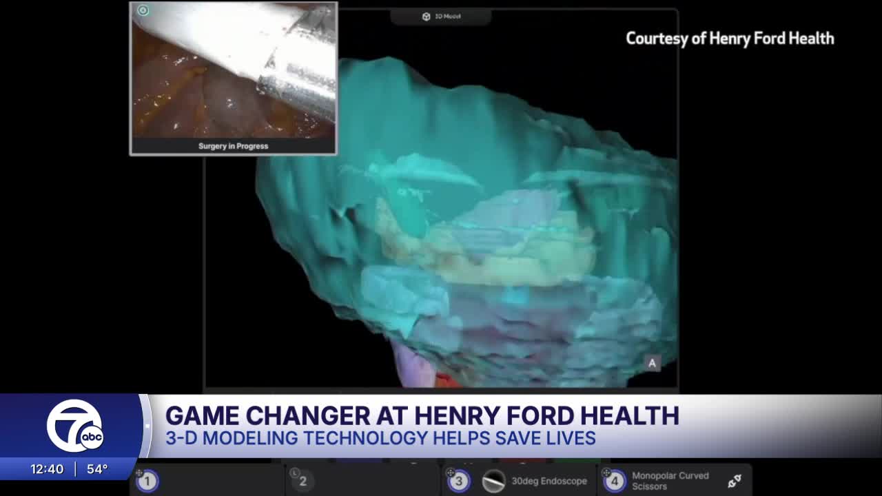
That plan was built using a 3D model from Jim's own scans. For years, surgeons relied on flat, 2D images, mentally building a 3D picture in real time.
But in the operating room, that picture comes to life.

Dr. Craig Rogers, Head of the Vattikuti Urology Institute at Henry Ford Health, says that the 3D models allow him to see the tumor and surrounding structures in real time, manipulate it, strip away layers, and map out exactly how to remove the cancer while protecting what matters most.
"So the benefit is more precision," Dr. Rogers said. "When you see things better, you operate better. You take better care of patients."
"Do you wish you had this earlier?" I asked Dr. Rogers.
"I do, yeah, I'm glad I have it now," he replied.
Now, Dr. Rogers can compare, via side-by-side view, both the model and Jim's actual kidney during surgery, no longer having to step away to use the old 2D scan. Adding precision can be the difference between removing a tumor and removing an entire organ.
"I can rotate it around and have greater confidence that I'm in the right place, that I am getting the tumor all out and saving the kidney," Dr. Rogers said.
It's a level of visualization only available at a handful of hospitals nationwide. But the impact isn't just in the operating room; it's something patients can see.
“When I saw that 3D colored image… even I could understand it… and it did build my confidence," Overbeck said.
It turns something complex into something clear. And in Jim's case, that clarity helped guide a plan to remove the tumor and save the kidney.
"He has two kidneys, and he's cancer-free," Dr. Rogers said.
It's a result that not that long ago may not have been possible.

"Last month I had cancer. Then I had the surgery, and now I'm cancer-free… and I'm back to playing hockey," Overbeck said.
He's spending time with his family, too. The work is so cutting-edge that Dr. Rogers is presenting Jim's case at an upcoming conference in Washington D.C., where thousands of urologists will study how 3D technology can help guide more complex kidney surgery.
